Skip to content

Precision dental care with digital X-rays

Advanced imaging for accurate diagnosis and effective treatment planning.

Girl in blue shirt standing in dental x-ray scanner.
Young person holding up x-ray of teeth.

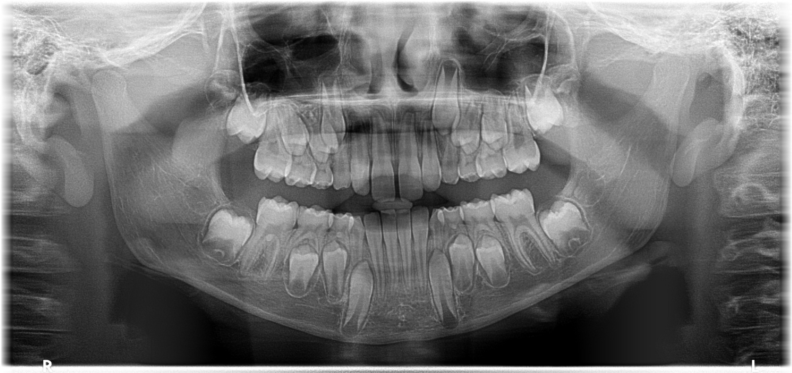
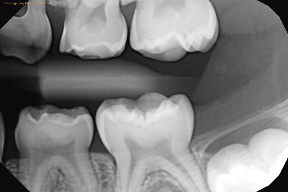
Benefits of digital x-rays

Digital X-rays are faster and contain less radiation than traditional radiographs, providing a safer and more comfortable experience. They offer immediate results, which means quicker diagnosis and treatment initiation.

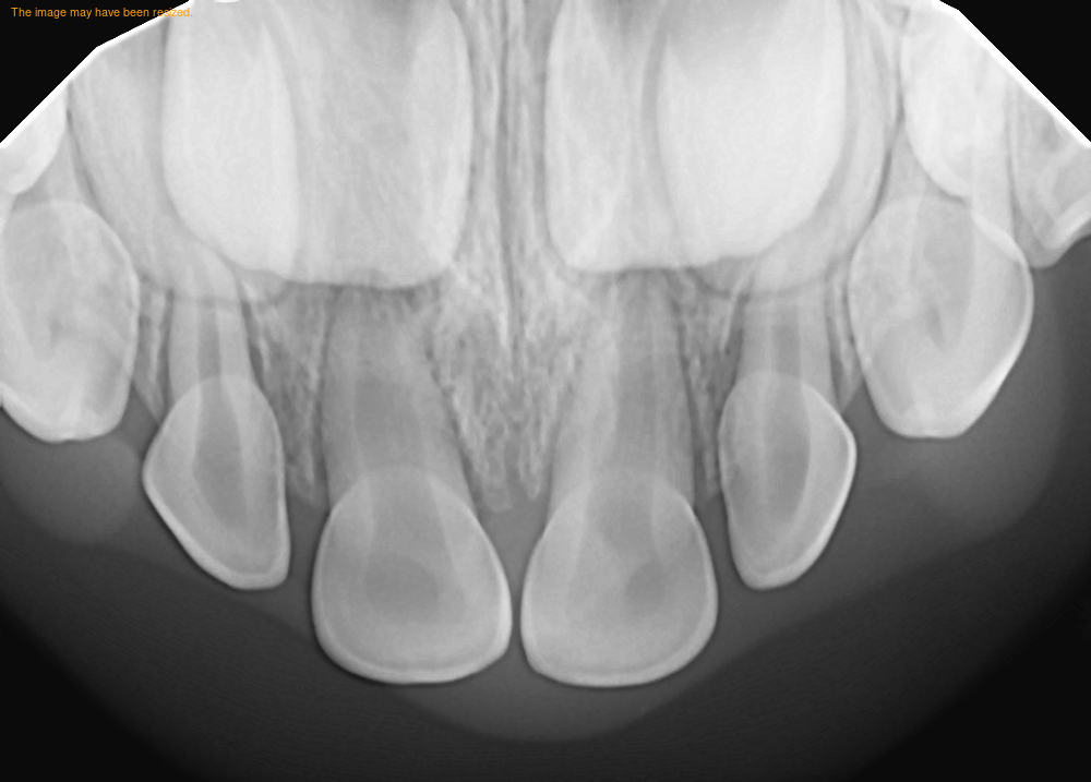
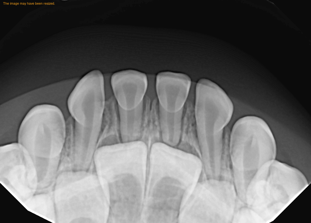
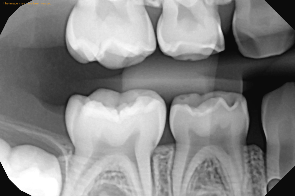
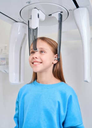
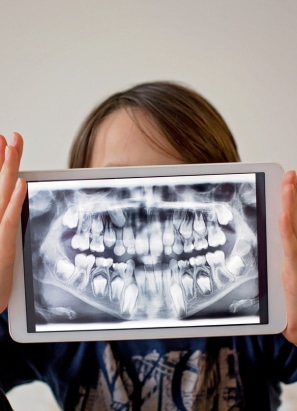
Integrating technology in dental care

Our dental practice integrates the latest in digital X-ray technology, ensuring that every diagnosis is based on the most accurate and comprehensive information available, leading to better dental health outcomes.

Revolutionizing dental diagnostics

X-rays are taken to gain information that a visual exam cannot provide. X-rays are needed to evaluate for cavities between the teeth, show unerupted, extra, or missing teeth, diagnose pathology, evaluate the results of an injury, and plan orthodontic treatment. Using digital x-rays means the amount of radiation that is detected is very small (less than flying in an airplane or spending one afternoon outside). Depending on your child’s age and cavity risk, our dentists will prescribe the appropriate radiographs per the American Academy of Pediatric Dentistry guidelines. These x-rays may include a panoramic, periapical, or bitewings films.

Our commitment to advanced diagnostics

We are dedicated to providing our patients with the highest standard of dental care, which includes utilizing advanced digital X-ray technology for all diagnostic needs, ensuring precision in every treatment plan we devise.

Types of digital x-rays

Comprehensive imaging for every need.


  • Upper and Lower Periapical (PA): Targets specific teeth for close-up views, identifying developing teeth, cavities, and root health.
  • Bitewing: Detects decay between development of teeth.
  • Panoramic: Offers a complete view of the mouth, showcasing all the teeth, jawbone (maxilla and mandible), and sinus areas in one image.
  • 3D Imaging: Provides three-dimensional views for complex diagnoses, treatment planning, and orthodontics.
  • Cephalometric Projections: Shows side views of the face and skull, used in orthodontic planning and TMJ assessments.